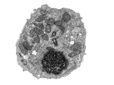
A Generalised Animal Cell as observed under an Electron Microscope

Some features common to animal cells. (Reproduced by permission of Photo

A Generalised Animal Cell as observed under an Electron Microscope

Alll about Elodea. elodea_cells.jpg

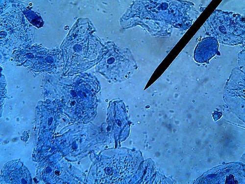
Each of these epithelial cells was examined under the microscope as students

Once you have a microscope, put a plant cell under

A Generalised Animal Cell as observed under an Electron Microscope.

The microscope has been focused to show the topography of the pellicle,

observe cheek cells under the microscope

Cells under microscope Foto sin derechos de autor

A Generalised Animal Cell as observed under an Electron Microscope.

Staining for tartrate-resistant acid phosphatase of osteoclast under light

Examine the oral surface of a sea star under a dissecting microscope and

Culture of cervical cancer cells under microscope.

Below: Human cheek cells 100X. Click on the photograph to view an
/138_2_177_183html/Volume%2520138(2),%2520pp.%2520177-183_img_3.jpg)
For each experiment at least 3 areas under microscope (approximately 200

Parts of a Typical animal cell ; 1.The network in resting stage of the
Boon Wee looked through a microscope to study the structure of a cell.

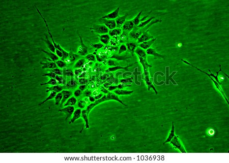
stock photo : Animal cells in culture viewed under an inverted microscope.

Rabies, seen here under a microscope, is an often fatal viral disease that
